How I Found Shenzhen Bay Laboratory
This summer, I saw the entire process through, from the first thought of applying for a research internship to finally working in a lab. A school email circulated the call for high school internships at the Shenzhen Medical Academy of Research and Translation, and my senior Lucy specifically pointed the program out to me — though I’d already had my eye on their high school research training opportunities for a while.
The Application Process
The application was anything but smooth. At first, I stared at the 241 people in the WeChat group and then realized there were only about 20 PIs (Principal Investigators), each taking just one or two students — the pressure was real. I talked it through with classmates who were also applying, and we ended up coordinating so we’d each target different labs.
Once the PI profiles went up in the group, I went through each one’s research area and weighed them against my two previous research experiences. Dr. Wang’s work stood out immediately — it aligned almost perfectly with what I’d already built. I’d previously done a gut microbiota survey on leopard cats, so I’d spent a lot of time with gut microbiome experiments and literature; and during my iGEM wall mold project, I’d dug into the literature on how post-disaster mold exposure affects respiratory health. Dr. Wang’s group happened to work on exactly these threads — gut microbiota interactions, infant nasal microbiome, and how environmental perturbations reshape the human microbiome. The fit was so clear I knew right then she was the PI I wanted.
Yet for my first choice, I didn’t go with the obvious fit. I picked Dr. Li, who works on biochemical proteins, because during iGEM I’d become fascinated with LC-MS and protein modeling — areas where I had no prior foundation and wanted to build one.
I want to take this chance to thank two of my teachers at school. When I was putting together the full application, both were enormously supportive. Mr. Sivanathan wrote my recommendation letter; Mr. Yates backed my application and helped arrange my school transcript. With their careful guidance, I wrote and proofread every piece of the application, submitted it on time, and waited with my fingers crossed for a chance to step into a real lab.
The application had been a rollercoaster, but I finally had my summer lab access — and I was ready to dive into this journey through the microscopic world of microbiota.
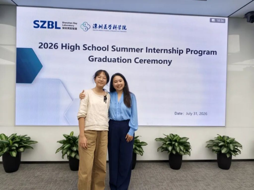
First Few Days: Stepping onto the Front Lines of Microbiome Research
On the afternoon of June 9th, the moment I put down my pen after finals, I could barely contain my excitement — summer freedom was finally here! I went home, packed, and on the morning of June 10th, I made my bed in the apartment and headed straight to the lab.
It was an early summer morning when I arrived at the Shenzhen Bay Laboratory. A scrolling digital screen by the elevators listed upcoming academic conferences, and the faint, crisp scent of an office building hung in the air. When I pushed open the door to Dr. Wang’s lab, the grad students and postdocs had just finished lab meeting and were still deep in discussion about plasmid construction logic. The rigorous, orderly atmosphere of real research hit me immediately.
The previous intern had reviewed the 2024 paper on WISH-tag technology and identified three core limitations of existing approaches: conventional 16S rRNA sequencing cannot distinguish subpopulations of the same bacterial strain with identical genotypes, making it difficult to precisely track the colonization dynamics of the probiotic EcN in the mouse gut.
Following the WISH-tag design principles in the paper, she selected five specific 40 bp barcode sets: W2, W26, W27, W28, and W29. Fusion PCR was used to amplify the 1500–1600 bp genomic integration fragment; after electrophoresis, the target band was precisely excised under UV light and the DNA was recovered and purified. EcN 1917 electrocompetent cells were then prepared, L-arabinose was added to induce λ-Red recombinase, and the five barcodes were site-specifically integrated into the bacterial malXY locus.
I took over the WISH-tag transformation plates she’d left behind, and the repeated colony PCR validation that followed turned out to be the most rewarding trial-and-error process of my summer. The first round yielded no positive bands at all. I broke down the variables one by one: standard purified water contained nucleases that degraded the template, and the initial identification primers lacked specificity.
After switching to nuclease-free water and resynthesizing improved primers, the second round of electrophoresis produced clean, sharp target bands. Those bright nucleic acid bands under UV imaging were the best possible reward for days of troubleshooting.
The mouse gavage in vivo tracing pilot was full of surprises. After separately administering W2 and W26 tagged strains, we collected mouse feces at 0.5, 1, 2, and 4 days, extracted genomic DNA, and performed qPCR quantification. The gut retention curves for the two strains were wildly different and had no reference value. We suspected the W2 mice were in poor health — their gut barrier was already compromised, which allowed the bacteria to proliferate massively after gavage.
There’s one story I won’t forget. Late one night, my teacher grabbed the wrong reagent — wash buffer instead of elution buffer — and the whole sample was compromised. Without hesitation, the two of us dug through the trash to retrieve the original sample tubes and redid the entire genome extraction from scratch. I’ll always remember calling my senior that night and him saying, completely unfazed, “It’s fine — we’ll just redo it.”
Failure is just part of doing biology, I suppose. This wasn’t my first time digging through a trash can, but it drove home the point that experiments go wrong — and when they do, you find a way to fix it. The one habit that saved us: keeping the original sample tubes tightly capped. Thankfully, nothing was contaminated.
To eliminate individual variability, we adjusted the protocol: all five WISH engineered strains were pooled and administered together by gavage, and the animal tracing pilot was repeated in full.
The second experiment was a gene functional validation built on dry-lab screening. The computational work had identified functionally relevant genes governing gut barrier translocation and systemic dissemination of Enterococcus gallinarum. Two distinct exposure groups showed divergent transcriptional patterns of sigma factor regulators: σ⁵⁴ was significantly upregulated in E. gallinarum, while σ⁷⁰ was markedly downregulated.
The experiment was designed to use CRISPR-Cas9 to construct a σ⁵⁴ knockout E. coli Nissle 1917 strain. Unfortunately, the plates showed purple mCherry colonies alongside both large and small white colonies. Colony PCR confirmed that the small white colonies were likely the correct ones — but all eight culture tubes stayed crystal clear. The inoculation had simply failed.
The CRISPR-Cas9–constructed σ⁵⁴ knockout E. coli Nissle 1917 was inoculated into the upper Transwell chamber to verify its ability to translocate from the apical side to the basolateral side. In the first pilot, the Transwell inserts suffered from medium overflow and massive cell detachment, with cells accumulating in the lower chamber.
For the second attempt, we adjusted the upper chamber volume to 150 μL and the lower to 600 μL, and optimized the collagen coating protocol. After the second seeding, microscopy revealed a uniform, intact cell monolayer adhering evenly across the membrane. After approximately two weeks of culture, we plan to measure the transepithelial electrical resistance (TEER).
Shadowing and Learning
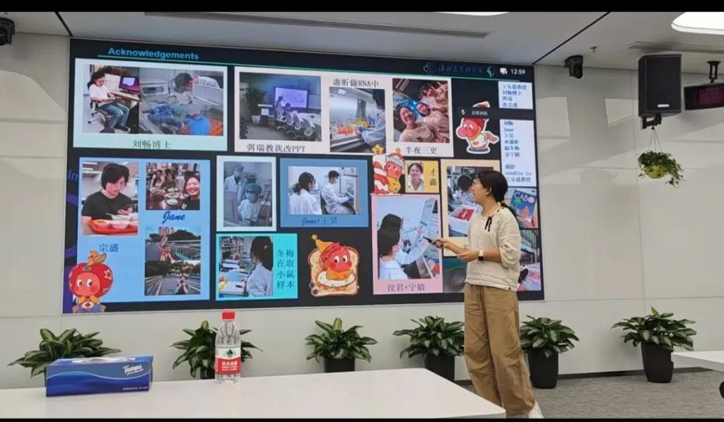
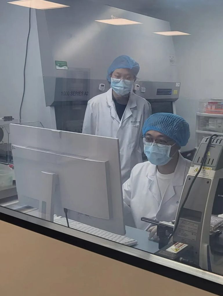
Beyond working on my teacher’s small wet-lab project, I shadowed other grad students and postdocs in the group, attended seminars, and sat in on flow cytometry training. I watched the entire wet-lab team process mouse samples together, working from daytime through midnight before they finally emerged from the lab — and then spending the next several days running flow cytometry with the orderly rhythm of an assembly line. It was even more impressive than last year, when I’d sonicated huge bags of protein all day in the iGEM lab — the experimental workflow here was simply more complex.
I also learned from a senior how to process mouse samples: adding grinding beads, running them in the homogenizer, extracting viral RNA, performing qPCR, and more.
This shadowing experience made me realize that before coming to the lab, I couldn’t even read many of the figures from wet-lab papers.
So over the summer I started studying immunology and infectious diseases, and once the school year began I picked up an excellent book a grad student had recommended — Immunobiology. I want to keep bringing infectious diseases, immunology, experimental principles, and figure-reading skills to my classmates — so this year I’m honored to serve as president of iHOSA, where I can pass on some of what I learned this summer to more students.
Downtime in the Lab
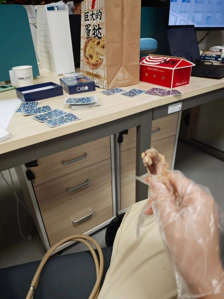
If there’s one thing two years of iGEM with friends gave me beyond lab skills and interview experience, it’s a habit of playing the card game CABO in our spare time. Playing with two underclassmen visiting from SCIE, friends in the group, and my senior — those are memories that won’t fade.
An Ending That Isn't Really an Ending
From receiving the internship call, to preparing my application with the help of two teachers, to embedding myself in the lab all summer — this journey pulled me out of the thin theoretical descriptions in textbooks and let me walk through a complete basic research cycle with my own hands. Nucleic acid bands glowing under UV gel, colonies slowly growing in the incubator, delicate intestinal epithelial cells under the microscope — countless microscopic images are etched into my memory.
Round after round of trial, error, reflection, and optimization didn’t diminish my love for microbiome research and synthetic biology — if anything, they showed me the true value of basic research: using rigorous, iterative engineering thinking to tackle real problems in human immunity and gut disease.
Dr. Wang’s team, with its broad research directions spanning mother-to-infant microbiota transmission, pathogen genome evolution, and environmental microbiota perturbation, has also pointed me toward a long-term path for further study. In the future, I hope to continue deepening my work at the intersection of host–microbe interactions, using gene editing and microbiota tracing technologies to explore how gut microbes regulate human health — and to pioneer new approaches for chronic disease management and targeted probiotic therapies.
As I gently close the heavy lab notebook, the hum of the biosafety cabinet and the soft click of the pipette still feel vivid and alive. This summer, thanks to my senior’s introduction and the support of two teachers at school, I stepped into Dr. Wang’s group, kept searching in the microscopic world of microbiota, and — through experiment after experiment and reflection after reflection — came to understand the unique rigor and romance of the life sciences.
This notebook is far from closed — the lights of Shenzhen Bay Laboratory are waiting for my return.
- Article / Nicole Yu